Skin Conditions


Albinism

Visit the following blogs to read about families who have adopted children with albinism:

Elora at Last
All Things Colbry
Adoption Makes Six
Adopting Jia
Chi Town Chronicles
Hopeful Hearts, Waiting Arms
1 More Piece


The word “albinism” refers to a group of inherited conditions. People with albinism have little or no pigment in their eyes, skin, or hair. They have inherited altered genes that do not make the usual amounts of a pigment called melanin. One person in 17,000 in the U.S.A. has some type of albinism.

Albinism affects people from all races. Most children with albinism are born to parents who have normal hair and eye color for their ethnic backgrounds.


1979900_10154039138120790_2550098371340955473_o


Sometimes people do not recognize that they have albinism. A common myth is that people with albinism have red eyes. In fact there are different types of albinism and the amount of pigment in the eyes varies. Although some individuals with albinism have reddish or violet eyes, most have blue eyes. Some have hazel or brown eyes. However, all forms of albinism are associated with vision problems.


Resources:

Raising a Child with Albinism
National Organization for Albinism and Hyperpigmentation (NOAH)
Videos by NOAH
Bai China Yahoo! Group
Albinism Adoptive Families Facebook Group
How a Person with Albinism Sees (video)
Albinism in FRAME (video)

Read blog posts about Albinism on No Hands But Ours.


Congenital Nevus

Visit the following blogs to read about families who have adopted children with Congenital Nevus:

Growing Beards

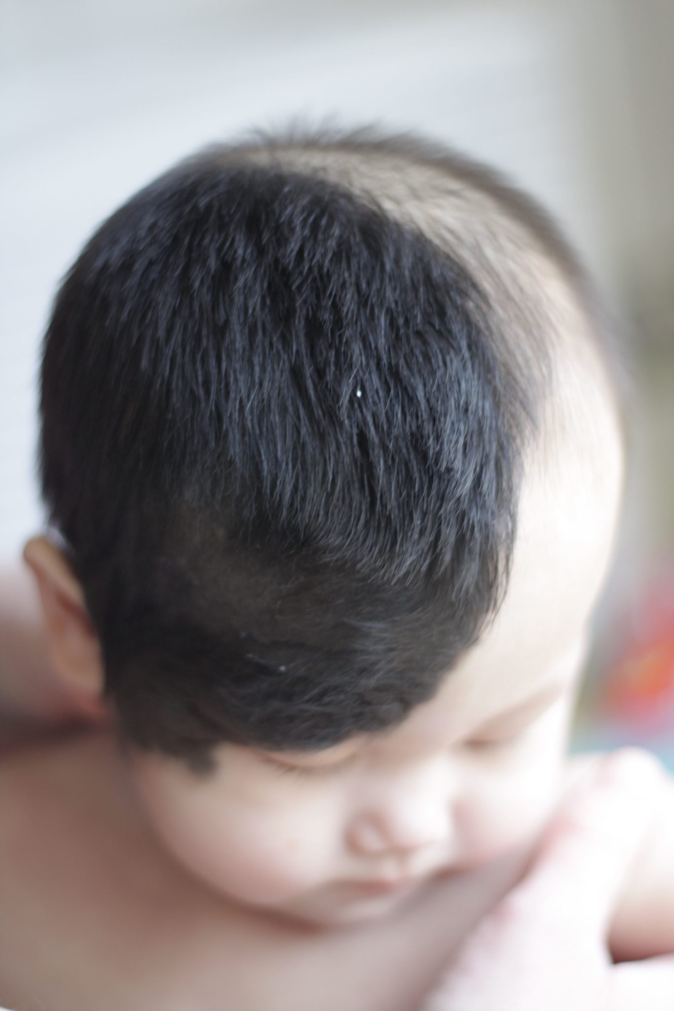
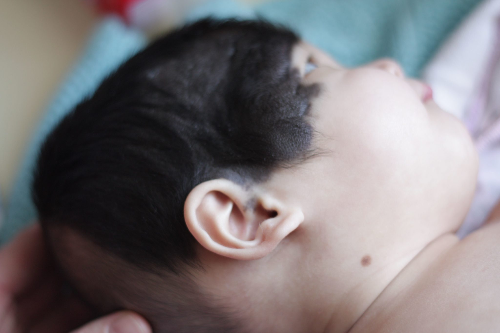
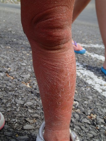
A Congenital Melanocytic Nevus can sound like an overwhelming special need. Simply, it is a birthmark (Nevus) present at birth (Congenital) that is made up of melanin or pigmented cells (Melancytic). Congenital Melanocytic Nevus (CMN) develops in the first trimester of fetal development and is NOT considered to be a hereditary or genetic condition. A nevus is often described based on the size of the birthmark: small, large, or giant. A large nevus covers more than 2% of the child’s skin, measures over 8”, would require more than one surgery to remove, or covers a large area of a body part such as an arm or leg. A large nevus is rare occurring in only 1 in 20,000 births. It is also very common to have “satellite” nevi, or smaller birthmarks, on other parts of the body in addition to the primary nevus.


rachels nevus


rachels nevus 2


There are some medical risks attached to this special need beyond the birthmark itself. An MRI is recommended to rule out a condition known as Neurocutaneous Melanocytosis (NCM). Individuals born with a large CMN have a 7% chance of having melanocytes (nevus cells) in their brain or spinal cord. More than 60% of those 7% of individuals with NCM, do not have any medical complications, even though an MRI conformed the presence of melanocytes in their nervous system. Complications from this condition may present as fainting, eyesight issues, hydrocephalus, or seizures.

Another risk is Melanoma, an aggressive form of skin cancer. Individuals born with a Congenital Melanocytic Nevus have a slightly elevated risk of Melanoma over the general population. It is estimated that a person born with a large nevus have a 2-5% chance developing Melanoma in their lifetime. An average person has a risk of Melanoma around 1% so current research estimates that an individual with a large, Congenital Melanocytic Nevus only has a slightly higher risk of developing this aggressive skin cancer. It is currently unknown if surgical removal of the nevus actually lowers the future risk of developing Melanoma.


Rachel 2014


With the unique needs and trauma involved in orphan care and adoption, it is important to evaluate the attachment and emotional needs of your child before committing to medical and surgical procedures that are non life-threatening. It is recommended to visit a pediatric dermatologist every 6 months for follow-up and evaluation.

A dermatologist can to determine if removal is necessary and other treatment options available. The decision to surgically remove a nevus is a highly debated, personal choice. If a family chooses to pursue removal, you would want to seek the opinion of a plastic surgeon in your area. Many Nevus families also chose to consult Dr. Bruce Bauer, Chicago, IL.

In recent years, the most common surgical removal option for a nevus is tissue expansion. Tissue expansion involves one surgery to place an expander (or multiple expanders) under the patient’s skin, expanding it over time with saline like a balloon, and stretching the skin to replace the nevus skin during a second removal surgery. It is fairly common for a patient to go through several rounds of expanders and removal surgeries to remove a large nevus.


Resources:
Nevus Outreach
Congenital Nevi
Giant Congenital Nevus
Giant Congenital Melanocytic Nevus
Nevus Love FB Support Group
Nevus Outreach FB Support Group

Read blog posts about Congential Nevus on No Hands But Ours.


Epidermolysis Bullosa

Visit this blog to read about a family adopting a child with Epidermolysis Bullosa:

From Life into Likeness4 Knights and the Princesses


Epidermolysis Bullosa (EB) is a very rare genetic connective tissue disorder. EB is actually a group of disorders that share a prominent manifestation of extremely fragile skin that blisters and tears from friction or trauma. Internal organs and bodily systems can also be seriously affected by EB. The list of secondary complications can be long and may require multiple interventions from a range of medical specialists. As of today, there is no cure or treatment. Daily wound care, pain management and protective bandaging are the only options available.


eb


The manifestations of EB, the symptoms the person suffers from, and the overall long term outcome on quality of life run the gamut from mild to devastatingly severe. There are many people who are diagnosed with milder forms of EB, which, while they can be extremely difficult and painful to live with, are not disfiguring or lethal. In these forms of EB blistering of the skin may be limited to the hands and feet which may not result in any scarring or loss of function. In more severe forms there is generalized blistering of the skin as well as injury to many internal organs and bodily systems. These more severe forms of EB result in disfigurement, disability and early death, usually before the age of 30. In fact, some forms of EB are lethal in the first few months of life (source).


Resources:
Mayo Clinic
Cincinnati’s Children
Hope, Wishes and Butterfly Kisses: EB Facebook Page
What is EB?
EB Resource: Sharing Our Story A New Beginning for Jie
The Long Hard Road with EB

Read blog posts about this special need on No Hands But Ours.


Ichthyosis

Visit these blogs to read about families who have adopted a child with Ichthyosis:

Preston Party of Six
Journey with Joy
Praying Gabby Home

Visit this blog to read about a family who has a child with Ichthyiois:

Blessed by Brenna


The ichthyoses (plural) are a family of genetic diseases characterized by dry, thickened, scaling skin. Because each form of ichthyosis is rare and there is an overlap of clinical features among disease types, the medical community disagrees about clear definitions and classifications of its many forms. Very rare forms of ichthyosis may also exist which are not fully described in the medical literature. Regardless of these difficulties, there are approximately 28 recognized forms of ichthyosis and related skin types. Most varieties of ichthyosis are relatively rare, affecting only one person in several tens of thousands. However, ichthyosis vulgaris is one exception; this form is estimated to affect one in every 250 people. Ichthyosis vulgaris is also the mildest form of ichthyosis and frequently goes undiagnosed or misdiagnosed as simply “dry skin.”


ichthyosis


Ichthyosis can be a disfiguring disease, and as such has numerous social and psychological implications. The more severe forms of ichthyosis can cause many other problems. When the skin loses moisture, it becomes dry, tight, and rigid. This rigidity makes moving uncomfortable as it can cause the skin to crack and break open. Extreme thickening of the skin on the soles of the feet makes walking difficult for many individuals, and cracking around the fingers can make even simple tasks difficult or painful. In some types of ichthyosis the skin is very fragile and will rub off with the slightest abrasion. Cracks and abrasions then leave the skin open to infections.

Severe scaling on the scalp may interfere with normal hair growth. Thick scales elsewhere can block pores, making sweating difficult and increasing the risk of overheating. Although the outer skin is thicker in ichthyosis, it is less effective in preventing water (and calorie) loss by diffusion across the surface of the skin. The rapid turnover of the outer layers of skin, in some forms of ichthyosis, also requires additional energy. Because of these greater energy needs, some children with severe ichthyosis may have trouble taking in enough calories to grow normally.

Some people with ichthyosis have trouble closing their eyes completely because the surrounding skin is so tight. This condition, called ectropion, causes the eyelids to turn outward, exposing the red inner lid and causing continuous irritation. If it is left untreated, damage to the cornea can develop, leading to impaired vision (source).


P5240145


Resources:
First Skin Foundation
Ichthyiosis.com

Read blog posts about Ichthyosis on No Hands But Ours.


© 2024 No Hands But Ours

The content found on the No Hands But Ours website is not approved, endorsed, curated or edited by medical professionals. Consult a doctor with expertise in the special needs of interest to you.