Urogenital


Anorectal Malformation

Visit these blogs to read about families who have adopted children with this special need:

A Homespun Heritage
Dragons and Elephants
Happy Go Luckie
La Dolce Vita
Love You Forever
There’s No Place Like Home


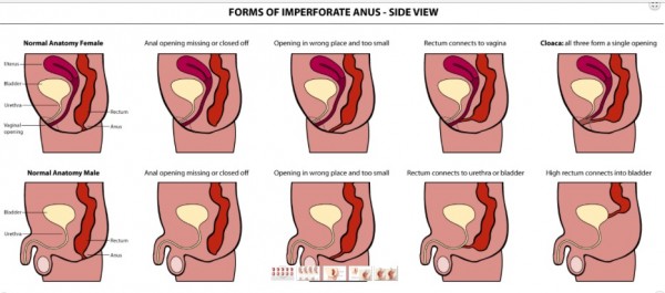
Anorectal Malformations (ARMs – sometimes referred to as anal atresia and imperforate anus) are defects that occur during the fifth to seventh weeks of fetal development. With these defects, the anus (opening at the end of the large intestine through which stool passes) and the rectum (area of the large intestine just above the anus) do not develop properly. Anorectal malformations affect 1 in 5,000 babies and is slightly more common in males. The exact cause of anorectal malformations is unknown.


9300_10202624138945784_2020286089502754266_n


During a bowel movement, stool passes from the large intestine to the rectum and then to the anus. Nerves in the anal canal help us sense the need for a bowel movement and also stimulate muscle activity. Muscles in this area help control when we have a bowel movement.

With an anorectal malformation, any of the following abnormalities can occur:
– The anal passage may be narrow or misplaced in front of where it should be located
– A membrane may be present over the anal opening
– The rectum may not connect to the anus
– The rectum may connect to part of the urinary tract or the reproductive system through a passage called a fistula, and an anal opening is not present


ARM image
Image credit


There are varying degrees of Anorectal Malformations (ARM) including high or low locations of the malformation. Children with low malformations are more prone to constipation (slow motility/hypomotile) while children with a shorter colon or a resection are more likely to have frequent, loose stooling (fast motility/hyermotile). A child born with an ARM defect will need surgery to correct the malformation and depending on how high the defect the child may need a temporary colostomy while the new anus heals. Some children from China come home with a colostomy and the remaining surgeries are done once home.

The surgery to correct an ARM is called the PSARP or posterior sagittal anorectalplasty. The PSARP approach was developed by Dr. Pena in 1980 and is also known as the Pena Pull-through. After the PSARP some children may require anal dilations to keep the new anus open.
While every ARM child’s condition is individual to them, many children can become “socially” continent through a bowel management program. A bowel management program can be tailored to each individual child and can include a daily stimulant laxative and/or daily enema. A few children are able to manage with diet alone but the majority of children will need a bowel management regimen.

When looking at the file of a child with an anorectal malformation it is important to remember that due to a lack of resources many ARM children are not properly maintained in China. These children may have had surgery, but present as incontinent. This could be due to surgery (which will require a re-do surgery once home), poor food choices (starches and other constipating foods), or lack of laxative/enema use. In addition, due to the chronic constipation found in many ARM children the child’s colon may be enlarged or stretched. ARM children’s colons will usually present as hypomotile (slow motility resulting in constipation) due to the original defect which affects the nerves and muscles. The stool moves very slowly eventually forming a stool ball making the child constipated. Over time, loose stool will begin leaking around the stool ball, and the child becomes incontinent due to the constipation. Children who have had their colons resected during a past surgery may present as hypermotile (fast motility resulting in frequent, loose stools). Many ARM children need either daily laxatives to help stimulate the colon to move, or daily enemas to fully empty the colon once a day. Once home, ARM children should be seen by a doctor well versed in ARMs to determine the best course of action. This plan includes having the child tested for VACTERL associated issues (tethered cord, heart, tracheoesophageal, renal/kidney and limb differences), a contrast enema, x-rays, and possibly a sedated exam to check the China repair and/or to better understand the child’s anatomy.

See also: Recto-vaginal fistula, Cloaca, Incontinence, and VACTERL.


DSC_0211


ARM Expert Doctors, Centers, and Hospitals in the US:
Cincinnati Children’s Hospital Colorectal Center, Cincinnati, OH
Nationwide Children’s Hospital Center for Colorectal and Pelvic Reconstruction, Columbus, OH
Nemours/Alfred I. duPont Hospital for Children, Wilmington, DE – Dr. Charles Vinocur
Children’s Hospital Colorado, Aurora, CO
Seattle Children’s Hospital, Seattle, WA – Dr. Jeffrey Avansino
Morgan Stanley Children’s Hospital, New York NY – Dr. Shumyle Alam, urology specialist

Resources:
Yahoo! Group – IA Adoptive Parent Support
Yahoo! Group – IA Parental Support
Facebook Group – Imperforate Anus/Adoptive Mamas (Please note this is for adoptive moms only)
Imperforate Anus – Cincinnati Children’s
Imperforate Anus – Nationwide Children’s
Posterior Sagittal Anorectoplasty (PSARP)/Pull-through
Overview of ARMs
Bowel Management Information (Nationwide Children’s)
Dr. Dickie (Cincinnati Children’s) open Q & A webinar
Urological Concerns in Anorectal Malformations part 1, part 2
Gynecologic Concerns in Girls with ARMs
Article by Dr. Dickie (Cincinnati Children’s)
MACE (Malone)/Cecostomy
Additional Information about the Malone antegrade continence enema (MACE) 
Children’s Hospital of Pittsburg
FAQs Bowel and Bladder Dysfunction adult adoptee, Alex Coe, answers FAQs
Pediatric Imperforate Anus Surgery
What Are Anorectal Malformations?
Bowel Management – an article by Drs. Levitt, Bischoff, & Pena
Pull-Thru Network
Continence Predictor Index


Read blog posts about Anorectal Malformations on No Hands But Ours.


Cloaca

Visit these blogs to read more about families who adopted daughters with this special need:

3 Lloyd Boys and Our Journey To Mayleigh Wei
Journey to Ava Mei

7

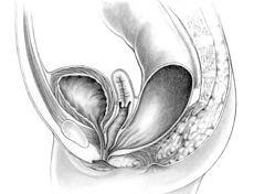
A cloaca is a type of anorectal malformation in which the rectum, vagina, and urethra open into a common area or single common channel. Cloacas occur in 1 in 20,000-25,000 live births. It is thought that this number may be higher. (Cloacas are commonly misdiagnosed as rectovaginal fistulas. This is concerning because this means that problems that are urological may go undiagnosed and untreated.) When diagnosing a cloaca there is the presence of a single perineal orfice. Only one opening is visible.

Initial treatment of the infant born with a cloaca is to divert the GI tract usually by colostomy, divert the urinary tract if necessary, and decompress the vagina if distended. After the infant recovers from the colostomy, the main repair of the cloaca is planned. This repair is called an anorectovaginourethroplasty. During this surgery the rectum, vagina, and urethra will be simultaneosly repaired and will be located as seperate structures. The colon will be pulled through to the place where the anal opening should be by creating an anus, the vaginal wall will be repaired if needed, and if located too high the urethra will be brought to the surface and repaired.

There are 3 main conscerns in regard to the child with a cloaca: urinary control, bowel control, and sexual function. If present, urinary incontinence can be well managed with medication and/or intermittent catheterization. Children with bowel incontienece can be socially continent using a bowel management program. Sexual function issues refer to menstration, intercourse, and obstetric issues.

cloaca

See also: Anorectal malformation, Recto-vaginal fistula, Incontinence, and VACTERL

Resources:
Nationwide Children’ Hospital: Cloacal Malformation
Cloaca Malformations
Gynecologic Concerns in Girls with ARMs
Cincinnati Children’s Hospital Colorectal Center, Cincinnati Ohio
Urological Concerns in Anorectal Malformations part 1, part 2
Nationwide Children’s Hospital Center for Colorectal and Pelvic Reconstruction, Columbus Ohio


Read blog posts about Cloaca on No Hands But Ours.


Cryptorchidism

Cryptorchidism is the most common congenital abnormality of the genitourinary tract. Cryptorchidism is the absence of one or both testes from the scrotum . Most cryptorchid testes are undescended, but some are absent (due to agenesis or atrophy).

10599550_10204598703076467_1248799023694951679_n

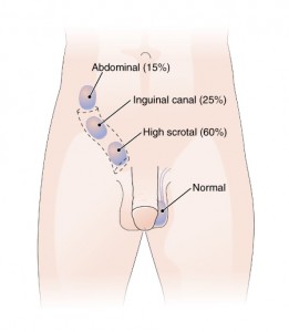
True undescended testes have stopped short along their normal path of descent into the scrotum. They may remain in the abdominal cavity or they may be palpable in the inguinal canal (intracanalicular) or just outside the external ring (suprascrotal). About 3% of full-term, and 30% of premature infant boys are born with at least one undescended testis. However, about 80% of cryptorchid testes descend in the first year of life (the majority within three months).

A testis absent from the normal scrotal position can be:
1. found anywhere along the “path of descent” from high in the posterior (retroperitoneal) abdomen, just below the kidney, to the inguinal ring;
2. found in the inguinal canal;
3. ectopic, that is, found to have “wandered” from that path, usually outside the inguinal canal and sometimes even under the skin of the thigh, the perineum, the opposite scrotum, or the femoral canal;
4. found to be undeveloped (hypoplastic) or severely abnormal (dysgenetic)

image
(Image Source).

About two thirds of cases without other abnormalities are unilateral; one third involve both testes. In 90% of cases an undescended testis can be felt in the inguinal canal; in a minority the testis or testes are in the abdomen or nonexistent (truly “hidden”).
Although cryptorchidism nearly always refers to congenital absence or maldescent, a testis observed in the scrotum in early infancy can occasionally “reascend” (move back up) into the inguinal canal. A testis which can readily move or be moved between the scrotum and canal is referred to as retractile.

Cryptorchidism is distinct from both monorchism, the condition of having only one testicle, and anorchism, the absence of both testes at birth (for more on anorchism see Disorders of Sexual Development).

Due to the high probability of self-resolution the primary management of cryptorchidism is watchful waiting. If the testis does not descended within the first year of life surgery (orchiopexy) is the most common treatment to bring the testes into the scrotum and is usually successful. Hormone therapy (B-HCG or testosterone) is sometimes attempted and occasionally successful.

Undescended testes are associated with reduced fertility, increased risk of testicular cancer. Undescended testes are also more susceptible to testicular torsion and infarction and inguinal hernias (Source 1 and 2).

Resources:
Medscape
Mayo Clinic
Boston Children’s


Hirschprung’s Disease

Visit these blogs to read about families who have adopted children with this special need:

Love, Laughter and Ladybugs
Roo’s Journey

Visit this blog to read about a family who has a biological child with this special need:

Sweet Baby Isaac


hirschprung


What is Hirschsprung disease (HD)?

Hirschsprung disease (HD) is a disease of the large intestine that causes severe constipation or intestinal obstruction. Constipation means stool moves through the intestines slower than usual.

Bowel movements occur less often than normal and stools are difficult to pass. Some children with HD can’t pass stool at all, which can result in the complete blockage of the intestines, a condition called intestinal obstruction. HD is diagnosed based on symptoms and test results.

People with HD are born with it and are usually diagnosed when they are infants. Less severe cases are sometimes diagnosed when a child is older. An HD diagnosis in an adult is rare.

Why does HD cause constipation?

The large intestine, which includes the colon and rectum, is the last part of the digestive tract. The large intestine’s main job is to absorb water and hold stool. The rectum connects the colon to the anus through which stool passes out of the body through the anus. People with HD have constipation because they lack nerve cells in a part or all of the large intestine. The nerve cells signal muscles in the large intestine to push stool toward the anus. Without a signal to push stool along, stool will remain in the large intestine.

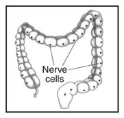
Healthy large intestine.
Nerve cells are found throughout the large intestine.

L_Intestine_Nerve_Cell1

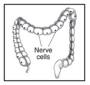
Short-segment HD.
Nerve cells are missing from the last segment of the large intestine.

L_Intestine_Nerve_Cell2

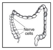
Long-segment HD.
Nerve cells are missing from most or all of the large intestine and sometimes the last part of the small intestine.

L_Intestine_Nerve_Cell3

How severe HD is depends on how much of the large intestine is affected. Short-segment HD means only the last part of the large intestine lacks nerve cells. Long-segment HD means most or all of the large intestine, and sometimes the last part of the small intestine, lacks nerve cells. In a person with HD, stool moves through the large intestine until it reaches the part lacking nerve cells. At that point, the stool moves slowly or stops, causing an intestinal obstruction.

What causes HD?

Before birth, a child’s nerve cells normally grow along the intestines in the direction of the anus. With HD, the nerve cells stop growing too soon. Why the nerve cells stop growing is unclear. Some HD is inherited, meaning it is passed from parent to child through genes. HD is not caused by anything a mother did while pregnant.

What are the symptoms of HD?

The main symptoms of HD are constipation or intestinal obstruction, usually appearing shortly after birth. Constipation in infants and children is common and usually comes and goes, but if a child has had ongoing constipation since birth, HD may be the problem.

Symptoms in Newborns:

— failure to have a first bowel movement within 48 hours after birth
— green or brown vomit
— explosive stools after a doctor inserts a finger into the rectum
— swelling of the belly, also known as the abdomen
— excessive gas
— bloody diarrhea

Symptoms in Toddlers and Older Children:

— not being able to pass stools without laxatives or enemas
— swelling of the abdomen
— excessive gas
— bloody diarrhea
— slow growth or development
— lack of energy because of a shortage of red blood cells, called anemia

How is HD treated?

HD is treated with surgery called a pull-through procedure. A surgeon removes the segment of the large intestine lacking nerve cells and connects the healthy segment to the anus. The pull-through procedure is usually done soon after diagnosis.

Most HD patients are treated using the pull-through, but sometimes an ostomy is needed. An ostomy allows stool to leave the body through an opening in the abdomen. Although most children with HD do not need an ostomy, a child who has been very sick from HD may need an ostomy to get better before the pull-through procedure. For ostomy surgery, the surgeon first takes out the diseased segment of the large intestine. The end of the healthy intestine is moved to an opening in the abdomen where a stoma is created. A stoma is created by rolling the intestine’s end back on itself, like a shirt cuff, and stitching it to the abdominal wall. An ostomy pouch is attached to the stoma and worn outside the body to collect stool. The pouch will need to be emptied several times each day.

Credit for above information and images

Hirschprung’s Disease doctors, centers, and hospitals in the US:
Cincinnati Children’s Hospital Colorectal Center
Nationwide Children’s Hospital Center for Colorectal and Pelvic Reconstruction
Seattle Children’s Hospital

Additional Resources & Support:
Hirschprung’s Disease FB group
Hirschsprung’s Disease Family Support page on FB
Nationwide Children’s ‘What is Hirschprung’s Disease
Nationwide Children’s Hirschprung’s Disease Info
Dr. Levitt (Nationwide Children’s) on Hirschprung’s Disease
Bowel Management by Dr. Levitt (Nationwide Children’s)
Cincinnati Children’s Hirschprung’s Disease Info
Hirschprung’s Disease – Mayo Clinic
NIH info on Hirschprung’s Disease
Dr. Levitt (Nationwide Children’s) YouTube Video on Bowel Management
PDF doc on Hirschprung’s Disease from Nationwide Children’s
Hirschsprings Disease, It’s All About Poop
Pull Thru Network
Hirschprung’s Disease Podcast – Dr. Levitt
MACE (Malone)/Cecostomy
Additional Information about the Malone antegrade continence enema (MACE) 


Hypospadias

Visit these blogs to read about families who have adopted children with his special need:

Faith, Family, Adoption
Following Where He Leads
One More Ladybug

hypospadias

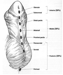
Hypospadias is a birth (congenital) defect in which the opening of the urethra is on the underside of the penis. The urethra is the tube that drains urine from the bladder. In males, the opening of the urethra is normally at the end of the penis. Hypospadias is a common birth defect occurring in approximately one in every 250 newborn boys; the cause of hypospadias is usually unknown.

Symptoms depend on the severity of the hypospadias. Usually, boys with this condition have the opening of the urethra near the tip of the penis on the underside, while in more severe forms the opening is in the middle or base of the penis. Rarely, the opening is located in or behind the scrotum. This condition may cause a downward curve of the penis during an erection.
Other symptoms include:
• Abnormal spraying of urine
• Having to sit down to urinate
• Foreskin that makes the penis looks like it has a “hood”

hypospadias image 2
(Image Source)

This problem is almost always diagnosed soon after birth during a physical exam. Imaging tests may be done to look for other congenital defects. Infants with hypospadias should not be circumcised. The foreskin should be kept for use in later surgical repair. Surgery is usually done before the child starts school.
10656338_10203934851445416_1765444447_n
Today, most urologists recommend repair before the child is 18 months old. Surgery can be done as young as 4 months old. During the surgery, the penis is straightened and the opening is corrected using tissue grafts from the foreskin. The repair may require multiple surgeries. Results after surgery are most often good. In some cases, more surgery is needed to correct fistulas or a return of the abnormal penis curve, and most males can have normal adult sexual activity (Source).

Resources:
Urology Health
Hypospadis information from Boston Children’s Hospital
Hypospadias information from Cincinnati Children’s Hospital
Hypospadias Repair Information


Read blog posts about Hypospadias on No Hands But Ours.


Incontinence

Visit these blogs to read about families who have adopted children with this special need:

There’s No Place Like Home
Feet 2 Our Faith
And Then We Found Kimi
Adopting Our Blessing

10660190_809466352432115_5819118919305539559_n

Incontinence is defined as a problem controlling urination or defecation. A person may be incontinent of urine, stool, or both.

Urinary incontinence

Urinary incontinence can be caused by UTI’s, obstructive sleep apnea, diabetes, kidney problems, constipation, structural problems in the urinary tract, or nerve problems. Structural problems in the urinary tract may cause leaking or increased bladder pressures that can be harmful to the kidneys. The problems could include bladder neck funneling or small bladder size. These things can be surgically corrected. Urinary incontinence caused by nerve problems is referred to as neurogenic bladder. This means that the person is unable to perceive bladder fullness and lacks the nerves to have coordinated bladder contraction and opening of the sphincter. Neurogenic bladders may be seen in children with spina bifida, anorectal malformations, spinal cord injury, and others. Treatments for urinary incontinence include intermittent catheterization and medications. Intermittent catheterization is done by placing a clean catheter through the urethra or stoma into the bladder to empty it and is usually done every 3-4 hours while awake. Another option for managing urinary incontinence is the Mitrofanoff appendicovesicostomy; a surgical procedure in which the appendix is used to create a small tunnel between the skin surface and the urinary bladder. The tunnel is used to empty the bladder using a catheter inserted into the bladder through the Mitrofanoff. Medications are also used to tighten the internal sphincter and to relax the bladder muscle to increase compliance. The goal for the urinary incontinent person is to be able to manage their incontinence and remain dry.

10681875_10204644642945789_1616719320_n

Fecal Incontinence

Fecal incontinence is when a person is unable to control their bowel movements. Like urinary incontinence this can be caused by a number of things including structural abnormalities, and nerve problems. Fecal incontinence is seen in children with anorectal malformations, spina bifida, spinal cord injuries, and others.

Initially it needs to be determined whether the person has “real” or “pseudo” incontinence. A child that acts as though they are incontinent but suffers for severe constipation/fecal impaction may regain control of bowel movements after the constipation is relieved. Real incontinence occurs when the muscles that surround the anorectal canal are altered and the sphincter that controls the anal opening is poor. Muscles need a correct nerve connection to function appropriately. A lack in these connections can occur in cases such as imperforate anus and spina bifida. This is also referred to as neurogenic bowel.

There are several ways to control fecal incontinence through what is known as bowel management. Fecal incontinence may be managed through diet, enemas, suppositories, laxatives, or with a Malone antegrade continence enema (also referred to as an appendicostomy, ACE, or MACE). The Malone requires surgery in which the appendix is used to create a small channel from the colon to the skin’s surface usually located in the belly button. This channel has a one way valve through which an enema solution is delivered to empty the bowels usually every day or every other day. The Malone is a good option for patients who use regular enemas to be socially continent but are looking for an alternate delivery system and eventually children can learn to do the flush independently. Bowel management is all about finding the perfect “recipe” of options to avoid accidents so that children can be socially continent.

10649530_10152418579193650_3739869863702025860_n

See also: Anorectal malformation, spina bifida

Resources:
AboutKidsHealth – information about the Mitrofanoff
MACE (Malone)/Cecostomy
Additional Information about the Malone antegrade continence enema (MACE) 
Link to Dr Levitt (Nationwide Children’s) YouTubeVideo on Bowel Management


Read blog posts about Incontinence on No Hands But Ours.

Recto-Vaginal Fistula

Visit these blogs to read about families who have adopted a daughter with this special need:

Henderson Home Online

1560771_10154645364705495_1321282013482682446_n

Recto-vaginal or Recto-vestibular fistulas are types of anorectal malformations found in females. Often times, this medical need is called imperforate anus or anal atresia with a recto-vaginal or vestibular fistula.

Both the recto-vaginal and recto-vestibular fistula are caused when the rectum does not connect properly to the outside of the body. Instead, a small passageway, called a fistula occurs, and stool passes through it and out through the vagina. The difference between these two conditions lies in the exit point: recto-vaginal has a higher exit point into the vagina while a recto-vestibular fistula has much lower exit point into the vestibular section of the vagina. These conditions are found when the child is examined at birth to check for proper opening and position of the vagina and anus.

ARM image

The corrective treatment for this condition includes either:
• the PSARP surgery which eliminates the fistula, reroutes the rectum through the sphincter muscle complex and creation of new anus without the use of a colostomy.
• Colostomy, anoplasty and reversal of colostomy which takes three separate surgeries.

Because this condition falls under ARM (anorectal malformation), children with a recto-vaginal or vestibular fistula, should also be screened for other VACTERL related conditions. The prognosis for children with recto-vaginal or recto-vestibular fistula is good since it is considered a low anorectal malformation. This means these children have a high chance of having bowel control and function.

See also: Anorectal malformation, VACTERL

Resources:
Mayo Clinic
Urological Concerns in Anorectal Malformations part 1, part 2
Cedars Sinai
Gynecologic Concerns in Girls with ARMs

Read blog posts about Rectovaginal or Rectovestibular Fistula on No Hands But Ours.


Underdeveloped/Ambiguous Genitalia

Read these posts about families who have adopted a child with this special need:

Our son’s Non-Special Need
Welcoming the Unknown

13606_10202827418941838_3717958779234951699_n

Disorders of Sex Development (DSD) are defined by the medical community as “Congenital conditions in which development of chromosomal, gonadal or anatomic sex is atypical.” DSD is an umbrella term covering a wide variety of conditions in which sex develops differently from typical male or typical female. Hermaphrodite, Pseudo-hermaphrodite and Intersex are considered outdated and inaccurate terms, however some in the medical community may still use them.

One of the most common concerns when considering a child with a DSD is what their future will look like. Many, many adults with DSD’s are happily married. Whether a person will be able to have biological children depends on their particular DSD, but remember, adoption is always an option. DSD’s can seem scary and overwhelming but they are manageable and more common that you think.

It is estimated that 1 in every 2000 live births results in a child whose genitals are atypical enough to make the sex unclear. However, it is also estimated that as many as 1 in every 100 live births results in a child with some form of sex anomaly.

When reviewing a child’s file their special need may be referred to as “Hermaphroditism”, ambiguous genitalia, vaginal atresia, micro-penis and so on. It is nearly impossible to know which DSD your child has based on those terms alone. Genetic testing in country is usually a simple 5 cell test to determine male-XY or female-XX genotype. That test alone, however, is not the determining factor in a child’s identity or phenotype. The presence of a Y chromosome does not make the child male any more than the absence of a Y chromosome make them female.

In most cases a child will have male-typical XY or female-typical XX sex chromosomes with varying degrees of atypical genital development. This can result in ambiguous genitalia or an appearance of the opposite sex. In some cases children will have a combination of sex chromosomes resulting in XXY, XX/XY, XO/XY and so on. With so many different genetic possibilities it is nearly impossible to name all of the known conditions that result in a DSD.

Two of the most common male-XY DSD’s are Complete Androgen Insensitivity Syndrome (CAIS) and Partial Androgen Insensitivity Syndrome (PAIS). CAIS and PAIS are caused by mutations in the AR gene. This gene provides instructions for making a protein called an androgen receptor. Androgen receptors allow cells to respond to androgens, or hormones (such as testosterone), that direct male sexual development. Mutations in the AR gene prevent androgen receptors from working properly, making cells less responsive to androgens or preventing cells from using these hormones at all. Depending on the level of androgen insensitivity, an affected person’s sex characteristics can vary from mostly female to mostly male. Most people with CAIS are raised as daughters. Those with PAIS will identify as male or female depending on the amount of androgen insensitivity.

5-alpha-reductase deficiency (5-ARD) is caused by mutations in the SRD5A2 gene preventing steroid 5-alpha reductase 2 from effectively converting testosterone to DHT in the developing reproductive tissues. As such, this condition affects male sexual development before birth and during puberty. Like CAIS and PAIS, people with this condition are genetically male and have gonads (testes). Their bodies, however, do not produce enough of a hormone called dihydrotestosterone (DHT). DHT has a critical role in male sexual development. A shortage of this hormone disrupts the formation of the external sex organs before birth resulting in the appearance of ambiguous or female genitalia. During puberty, the testes produce more testosterone. Researchers believe that people with 5-ARD develop secondary male sex characteristics in response to higher levels of this hormone. 5-ARD is quite rare but it is estimated that about half of people with this condition will adopt a male identity as an adult regardless of a childhood female identity. Gonad removal and feminizing surgery is usually done for those wishing to remain female.

Some conditions cause ambiguous genitalia but are not considered a Disorder of Sex Development. Congenital Adrenal Hyperplasia (CAH) is one such condition. While manageable with daily medication, left untreated some forms of CAH can be life threatening. CAH prevents the adrenal glands from producing cortisol. The pituitary gland attempts to compensate for the lack of cortisol by producing more and more ATCH. ATCH is converted by the adrenal glands into androgens resulting in a masculinizing effect on the genitals. As such, female genitalia is more often affected than male.

When adopting a child with a DSD it is critical to have a qualified and experienced team of specialists at your disposal. Most major children’s hospitals will have a specialized group for just such needs. Your child will need to be assessed shortly after coming home by a pediatric gynecologist, urologist, endocrinologist and geneticist to determine what DSD your child has, as well as the best course of action. The comprehensive testing can be a slow process taking 6 months or more. Often these specialized teams will include a psychologist to help the parent (and child) understand and work through the emotional side of DSD’s.

Sometimes corrective surgeries are necessary as is hormone replacement therapy. An experienced urologist who does a minimum of 3-4 specialized corrective surgeries a year is a must. The surgery should leave the head of the phallus intact preserving all of the nerve endings. Hormone replacement is critical for longevity and health if the child does not produce them on their own. This is administered by an endocrinologist following your child’s growth patterns. Working closely with your child and team of doctors you will be able to make the very best choices for your son or daughter’s future.

Resources:
Accord Alliance (this site has an excellent free printable handbook for parents)
DSD Families
Diagnosis and Treatment of the Androgen Receptor
Rainbow Kids: Our Son’s Non-Special Need
Steroid 5-alpha-reductase-2 deficiency
Rainbow Kids 

Read blog posts about Underdeveloped/Ambiguous Genitalia on No Hands But Ours.


VACTERL

Visit these blogs to read about families who have a child with this special need:

VACTERL SCHMACTERL
La Dolce Vita
Stops Along the Journey

10682596_10204863389249603_36156755_o

VACTERL Association (sometimes referred to as VATER syndrome/association) is an acronym used to describe a series of non-random birth defects which occur together.

V stands for vertebral anomalies

A stands for imperforate anus or anal atresia

C stands for cardiac (heart) defects. The most common heart defects seen with VACTERL association are ventricular septal defect (VSD), atrial septal defects (ASD), and tetralogy of Fallot.

TE stands for tracheal-esophageal fistula (TEF) or esophageal atresia (EA). Tracheal-esophageal fistula is an abnormal connection between the trachea (wind pipe) and the esophagus. Esophageal atresia occurs when the upper part of the esophagus does not connect with the lower esophagus and stomach.

R stands for renal or kidney anomalies.

L stands for limb abnormalities which can include a displaced or hypoplastic thumb, extra digits (polydactyly), fusion of digits (syndactyly), and forearm defects such as radial aplasia.

Babies diagnosed as having VACTERL association usually have at least three or more of these individual anomalies. Due to the wide range of manifestation of VACTERL association the exact incidence within the population is not known, but has been estimated to occur in one in 10,000 to 40,000 newborns. The reason it is called an association, rather than a syndrome, is that the complications are not pathogenically related but do tend to occur together more frequently than expected by chance. VACTERL association can be linked to other similar conditions such as Klippel Feil and Goldenhar Syndrome including crossovers of conditions; however, no specific genetic or chromosomal abnormalities have been identified with VACTERL association. Affected individuals may have additional abnormalities that are not among the characteristic features of VACTERL association (credit 1 and 2).

10500519_10203547011928638_7847613289406933278_n

See also: imperforate anus, heart defect, tracheoesophageal fistula, tethered spinal cord.

Resources:
NIH VACTERL Association
VACTERL Network
Orphanet Journal of Rare Diseases – VACTERL/VATER Association
VACTERL Association Facebook Group
VACTERL Association Support Group (UK based – alternate spelling of esophageal)


Read blog posts about VACTERL on No Hands But Ours.


© 2024 No Hands But Ours

The content found on the No Hands But Ours website is not approved, endorsed, curated or edited by medical professionals. Consult a doctor with expertise in the special needs of interest to you.