Orthopedic


Amniotic Band Syndrome

Visit these blogs to read about families who have adopted a child with amniotic band syndrome:

My Amazing Journey
Red Thread Joy
Table for Seven
Unexpected Paths
Popsicles and Popcorn


10668056_10105328091179444_1899625356_o


One of the primary causes of deformities of the fingers and toes found in children in China is a result of Amniotic Band Syndrome (ABS). ABS results in congenital birth defects believed to be caused by entrapment of fetal parts (usually a limb or digits) in fibrous amniotic bands while in utero.

Some common deformities that result from ABS are:

    • missing digits
    • missing limbs
    • webbing between fingers/toes
    • fused fingers/toes
    • clubfoot (sometimes)
    • cleft lip/palate (rarely)

This is a birth defect that in no way impairs the cognitive function of the baby; it is strictly a physical issue. In some cases, there is a need for surgery to create separation of the webbing to increase use and mobility. In the cases where a limb is missing, often no surgery is needed. For clubfoot, it is treated as any other child born with this issue, either by serial casting, surgery or the help of Ankle-Foot Orthoses (AFO).


abs2


Resources:
Amniotic Banding Syndrome
Children’s Hospital of Wisconsin
Scottish Rite: Hand Disorders
Scottish Rite Hand Camp
Scottish Rite Hand Support Group


Read blog posts about Amniotic Banding on No Hands But Ours.


Arthrogryposis

Visit these blogs to read about families who have adopted a child with arthrogryposis:

Peaches and Pandas
Breezy Acres Farm
Feet 2 Our Faith

10658929_10152285158432765_7828465579680282482_o

Arthrogryposis is a term used to describe a number of rare, non-progressive conditions characterized by stiff joints and abnormal muscle development. It is also referred to as arthrogryposis multiplex congenita or amyloplasia.

The exact cause of arthrogryposis is unknown, though a number of different theories have been proposed. Some believe that arthrogryposis is caused by mechanical obstructions to intrauterine movement during pregnancy. Others believe that it may be a result of an early viral infection during development. Still others believe that arthrogryposis is the result of failure of the central nervous system and/or muscular system to develop appropriately. Arthrogryposis is not thought to be a genetic or hereditary condition (Source).

10660990_10204644638625681_1805951232_n

Resources:
Boston Children’s Hospital
AMC Support
Introduction to Arthrogryposis part 1 (YouTube), part 2, part 3
Yahoo Artrogryposis Group
Rainbow Kids
3D Printed “Magic Arms” (video)
AMC Support Facebook Group
Arthrogryposis Facebook Group


Read blog posts about Arthrogryposis on No Hands But Ours.


Clubfoot

Visit these blogs to read about families who have adopted a child with clubfoot:

Ni Hao Y’all
Live Your Life
Made in China With Love
Casting a Stone
Tired Mama
An Unexpected Blessing
Surpassing Greatness
Greatly Blessed

image

Clubfoot, also known as talipes equinovarus, is a common congenital birth defect present in approximately 150,000 newborns worldwide every year. It affects boys slightly more frequently than girls and bilateral clubfoot (when both feet are affected) occurs in 30-50% of cases. The cause of clubfoot is largely unknown, but environmental factors and genetics are believed to play a part. When a child is born with the condition, the affected foot is turned upward and inward. If left uncorrected, the child would walk on the side or top of the foot. The goal of treating the clubfoot is to achieve a foot that looks and functions as much like a normal foot as possible. Most doctors agree that the initial treatment should be non-operative. Unfortunately, many doctors are not correctly trained in the Ponseti Method of serial casting, and surgery becomes too often the rule, instead of the exception.

What is the Ponseti Method of treatment?

Dr. Ponseti began developing a method to correct clubfoot without surgery in 1948. Dr. Ponseti passed away in October of 2009, at the age of 95. He continued to treat children with clubfoot at the University of Iowa Children’s Hospital, until just months before he passed away.

Ponseti’s method involves a series of plaster casts, applied from toe to groin, changed every 5 to 7 days. The doctor gently manipulates the bones of the foot in a specific order. The cast holds the foot in the new position, gently stretching the tendons and ligaments. With each cast change, the foot is manipulated in small increments, until the last cast achieves full correction. Most children with clubfoot require 5 – 7 casts and only in atypical cases are more casts necessary. For a child treated from birth, no more than 9 casts are required to achieve full correction.

A percutaneous tenotomy is required in approximately 80% of patients to lengthen the Achilles’ tendon to complete the correction. This procedure can be done in an office setting with local anesthesia. It involves a small poke with a tiny scalpel in the child’s heel and requires no stitches to close the wound. After the tenotomy, the final cast is applied, which stays on for 3 weeks in order for the tendon to regenerate to the proper length. When the last cast is removed, the child begins wearing a brace that holds the corrected foot in a stretched position, essentially ‘retraining’ the body to recognize the new, corrected, foot alignment; similar to the use of a retainer after orthodontic braces are removed.

The brace consists of two shoes, connected by a bar. The brace is worn for 23 hours a day for the first 3 months. Over time, the daily bracing hours are gradually reduced. By the time the child is walking, the bracing hours are reduced to bed time only. The brace is worn at night until approximately 5 years of age. The tendency for the foot to relapse remains active for years, but diminishes over time as the child grows. The exact causes of relapse are still being studied. At this time there are no criteria to determine whether or not a child’s foot is prone to relapse. Wearing the brace to keep the foot stretched gives the child the best chance to avoid relapse. After they are released from the brace wear, there are no special requirements and, assuming the child has no additional health issues, they may pursue an active lifestyle with no restrictions. When applied by a skilled physician, Ponseti’s method is successful in achieving complete correction in nearly 100% of patients with congenital clubfoot.

Why is serial casting better than surgery?

The Ponseti method has many advantages over surgical reconstruction. The first consideration is how much easier gentle manipulations and serial castings are for the patient to endure. Casting is not painful and often the child watches curiously as the foot is gently manipulated and as the plaster casts are applied. Secondly, surgery often makes the clubfoot ‘look’ correct, but internally the components of the foot and leg have been weakened. Excessive scar tissue, stiffness and limited motion can be effects of surgery, occurring early in the patient’s life and lasting a lifetime. This leaves the patient with a somewhat normal looking foot, but with potentially debilitating foot pain. Patients who have had their clubfoot reconstructed surgically often require additional surgeries over time; which can lead to more scar tissue and complications.

1924370_10152705719724784_1338552708609345009_n

Resources:
Ponseti Interational
University of Iowa
Shriners Hospitals
Children’s Hospital of St. Louis
Yahoo! Group – No Surgery 4 Clubfoot
Yahoo! Group – Clubfoot Adoption
Facebook Group – Clubfoot is Treatable
Facebook Group – Clubfeet
Facebook Group – Thank You, Dr. Dobbs


Read blog posts about Clubfoot on No Hands But Ours.


Dwarfism

Visit the following links to read about families who have adopted a child with dwarfism:

Bringing Home Holland
Full Plate Mom
One Love, One Family
Redeemed Mama
Roll You Home
The OASIS – Adopting His Children
Building our Family by Birth and Adoption
Little People, Big Decisions
Ni Hao Y’all


unnamed


Dwarfism is a genetic condition that occurs in approximately 1 in 10,000 births. Achondroplasia is the most common form of dwarfism, representing about 70% , however there are over 200 different recognized forms of dwarfism. It is often characterized by disproportionately short arms and legs, but some forms result in proportionate limbs. It might not be possible to determine the exact form of dwarfism a child has before he or she is adopted, therefore parents should be prepared for some unknowns in regard to an exact diagnosis.

Most people with dwarfism have equal intelligence and life span as their average height peers. Orthopedic complications are not uncommon in people with dwarfism, however most people with dwarfism enjoy good overall health. Some minor surgeries (ear tubes, adenoid and/or tonsil removal) are fairly common. People with dwarfism are able to do most things that their average height peers can do, although they may do it in a different way. Children can participate in many sports, although full contact sports such as football are not advisable. DAAA (Dwarf Athletic Association of America) organizes sports events for individuals with dwarfism at the national LPA (Little People of America) convention each year.


10644874_10202971968360707_8819196567222616729_n (2)


Resources:
Little People of America
LPA Blog
POLP (parents of Little People) on Facebook
PALS (parents of adopted Littles)
Rainbow Kids


Read blog posts about Dwarfism on No Hands But Ours.


Limb Difference

Visit these blogs to read about families who have adopted children with limb differences:

The Woller World
Hope 4 The Wounded
Learning to Fly with Chopsticks
Lila’s Turn
Scary Mommy
Greatly Blessed
Surpassing Greatness

DSC01279

The term, Limb Differences, is used in reference to the congenital (something a person is born with) absence or malformation of limbs. Some limb differences may be acquired as the result of an injury or disease that requires amputation. The causes of congenital Limb Differences are frequently unknown.

Challenges

A child who has an acquired limb difference through surgical amputation is more likely to feel a profound and deeply personal sense of loss. The child must adjust to a different way of living. This takes emotional and physical adjustments. Just as different amputation levels place different demands on a person, the demands resulting from amputation or a congenital difference are different.

A child’s emotional reaction to limb differences may vary. Some medical literature states that children who are born with a limb difference do not feel a sense of loss. Their body is the only body they have ever known. According to this point of view, it’s rare in the early years for a child who has a congenital difference to grieve. Still, as the child grows and becomes more socially aware, there can be a sense of loss or of being physically “different” from those who have fully functional limbs. This can then lead to frustration (“Why me?”) and some of the same feelings that occur while grieving.

Treatment

Infants and very young children with a lower-limb difference can better explore their world by wearing a prosthesis, as it will help that child get to a standing position more easily. Children limb differences affecting the arms often learn to compensate very early for the missing limb. With the love and support of a family, these children go on to live active, full lives (Source).

DSC05096

Resources:
Rainbow Kids
LimbDifferences.org
Yahoo! Group – Adopting Children with Limb Differences
Facebook Group – Limb Differences Red Thread China
Lucky Fin Project
Helping Hands Foundation
Living One Handed
Born Just Right
Camp No Limits
Super Hands
I-CAN International Child Amputee Network
Amputee Coalition
Shriners Hospitals


Read blog posts about Limb Differences on No Hands But Ours.


Osteogenesis Imperfecta

Visit these blogs to read about families who have adopted a child with OI:

Journey to Olivia
The O’Cain Crew
Adopting a Child with Osteogenesis Imperfecta

OI1

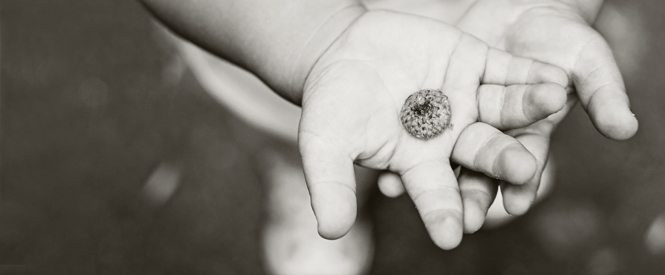
Osteogenesis imperfecta (OI) is a genetic disorder characterized by fragile bones that break easily. Also known as “brittle bone disease,” a person is born with this disorder and is affected through his or her lifetime. As a parent of a child/children with OI, our job is to encourage, empower, and equip them, and not succumb to the temptation to cover them in bubble wrap.

In addition to fractures people with OI may have muscle weakness, hearing loss, fatigue, joint laxity (loose and very flexible joints), curved bones, scoliosis, blue sclera (the whites of your eyes), dentinogenesis imperfecta (DI/brittle teeth), and short stature. Restrictive pulmonary disease occurs in more severely affected people.

OI is caused by an “error” or a mutation on a gene that affects the body’s production of the collagen found in bones, and other tissues. It is not caused by too little calcium or poor nutrition. Collagen act as the shock absorbers in your bones. Because the collagen is faulty, when the bones are bumped, there is nothing to cushion them, and so they break. Approximately 35% of children with OI are born into a family with no family history of OI. Most often this is due to a new mutation to a gene and not by anything the parents did before or during pregnancy.

OI is variable with 8 different types. The types range in severity from a lethal form to a milder form with few visible symptoms. The specific medical problems a person will encounter will depend on the degree of severity. A person with mild OI may experience a few fractures (there are some OI kids who play lacrosse!) while those with the severe forms may have hundreds in a lifetime (and spend their time in a wheelchair). The type of OI an adopted child has will probably not be known. Knowing the severity (mild, moderate or severe) of the child’s OI is helpful but may not be accurate if stated from the country of birth.

Diagnosis for OI is primarily based on signs seen in a doctor’s examination. When there is uncertainty about the diagnosis, it is best to consult a physician who is familiar with OI. Genetic testing is available to confirm a diagnosis of OI through collagen or gene analysis—a skin sample or a blood sample are used. While this is a nice thing to know, it is expensive and not necessary.

The OI type descriptions provide general information about how severe the symptoms probably will be. Health issues frequently seen in children and adults who have OI include:
• Short stature
• Weak tissues, fragile skin, muscle weakness, and loose joints
• Bleeding, easy bruising, frequent nosebleeds and in a small number of people heavy bleeding from injuries
• Hearing loss may begin in childhood and affects approximately 50% of adults
• Breathing problems, higher incidence of asthma plus risk for other lung problems
• Curvature of the spine

Doctors who see children and adults with OI include primary care physicians, orthopedists, endocrinologists, geneticists and physiatrists (rehabilitation specialists). Other specialists such as a neurologist may be needed. Most Shriners hospitals have seen OI patients. Of course the more they see the better informed they are in treating. In my (Jewel’s) experience they also do their best to assist you in being as independent as you are comfortable. In other words, if you can splint or wrap at home and don’t need to be seen except for extreme breaks, they can help you deal with that.

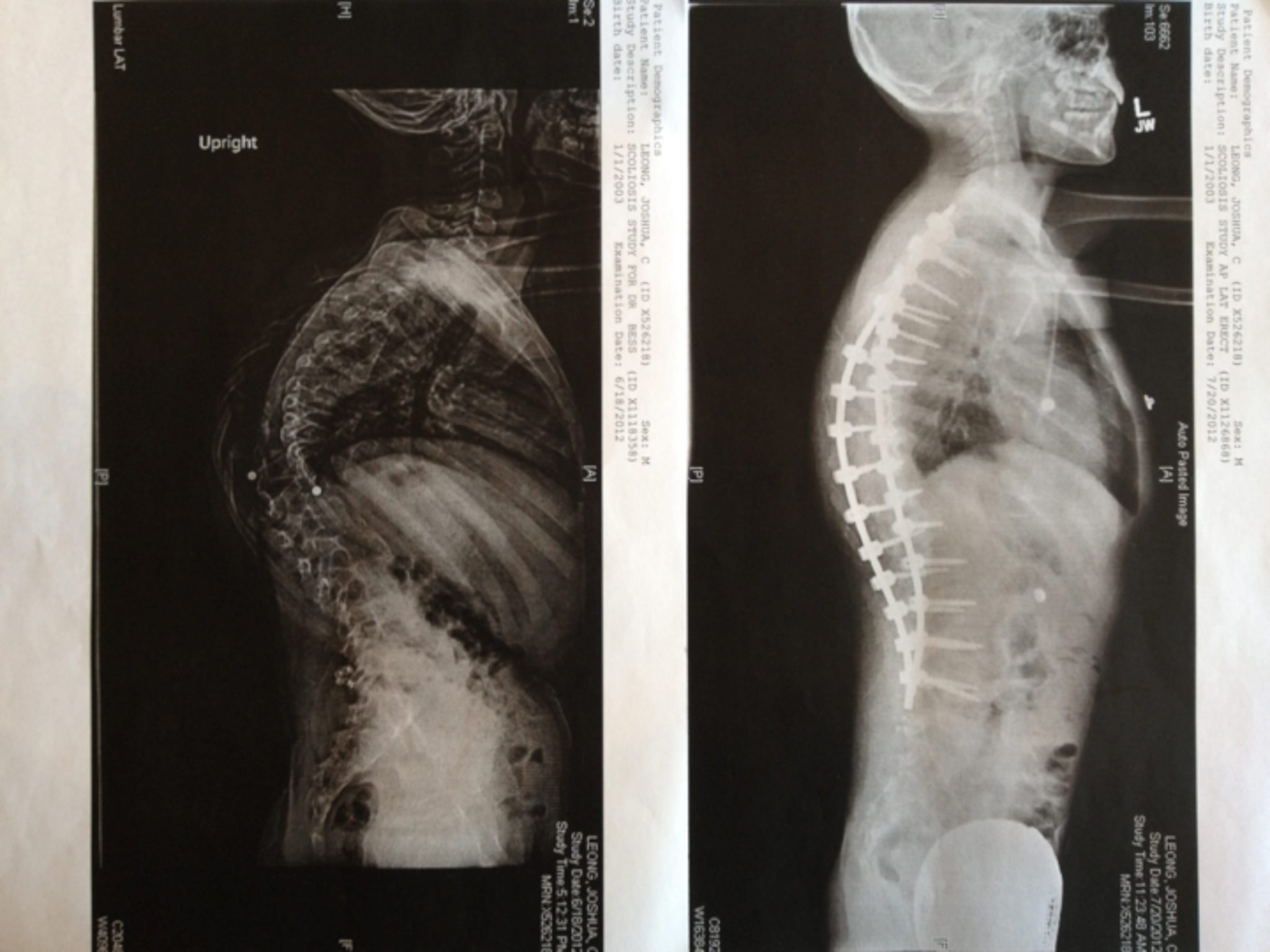
Treatments focuses on minimizing fractures, maximizing mobility, maximizing independent function and general health; physical therapy and safe exercise including swimming; casts, splints or wraps for broken bones; braces to support legs, ankles, knees and wrists as needed; orthopedic surgery, often including implanting rods to support the long bones in arms or legs; medications to strengthen bones; and mobility aids such as canes, walkers, or wheelchairs and other equipment or aids for independence may be needed to compensate for weakness or short stature.

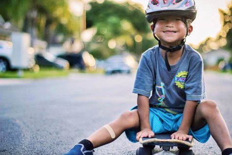
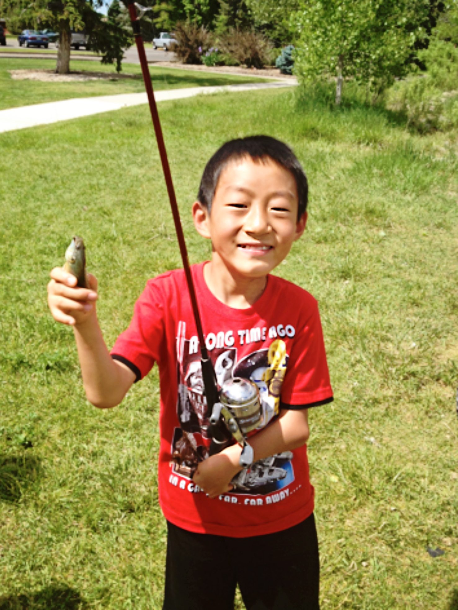
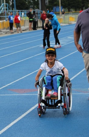
At this time, there is no cure. The prognosis for a person with OI varies greatly depending on the number and severity of symptoms. Life expectancy is not affected in people with mild or moderate symptoms, it may however, be shortened for those with more severe symptoms. The most severe forms result in death at birth or during infancy. Respiratory failure is the most frequent cause of death for people with OI, followed by accidental trauma. Despite the challenges of managing OI, most adults and children who have OI lead productive and successful lives. They attend school, develop friendships and other relationships, have careers, raise families, participate in sports (track, see below) and other recreational activities and are active members of their communities (Source).

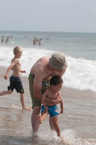
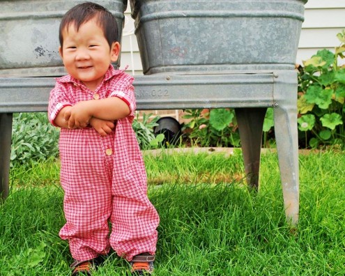
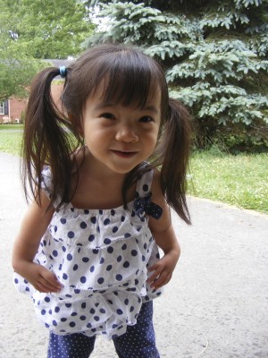
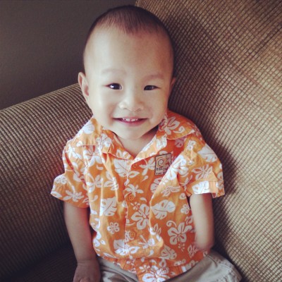
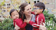
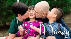
Note from Jewel, a mother who has adopted a daughter with OI (pictured below) –
“Personally, my daughter with OI is a joy to be around! She was adopted at 8 years old and is now 9. She expresses sorrow for her friend (who has yet to be adopted) who is afraid to stand on his own and perhaps walk. She understands the fear but she says, ‘I would try to walk!’ And she is trying now that her legs have rods and are straight, it won’t be long now. While she will probably never walk without some assistance, cane or walker, she will have the freedom to stand and walk as tolerated. She told me once that she would rather hurt herself and experience things than to just sit at home and do nothing except be “safe.” A family provides a stable, consistent environment of care for an adopted child with OI. If they did receive treatment, drugs or surgeries in their birth country, that treatment was probably not given as consistently as mom or dad will work to provide and would not be adequate care!”

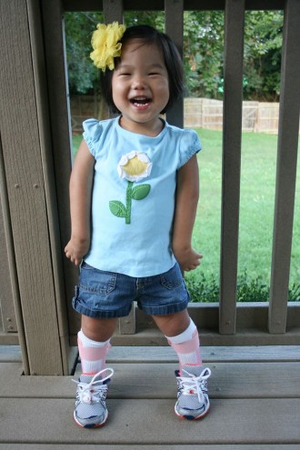
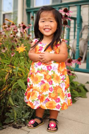
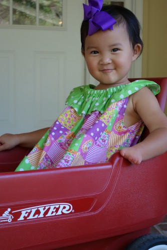
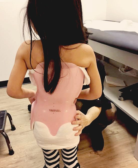
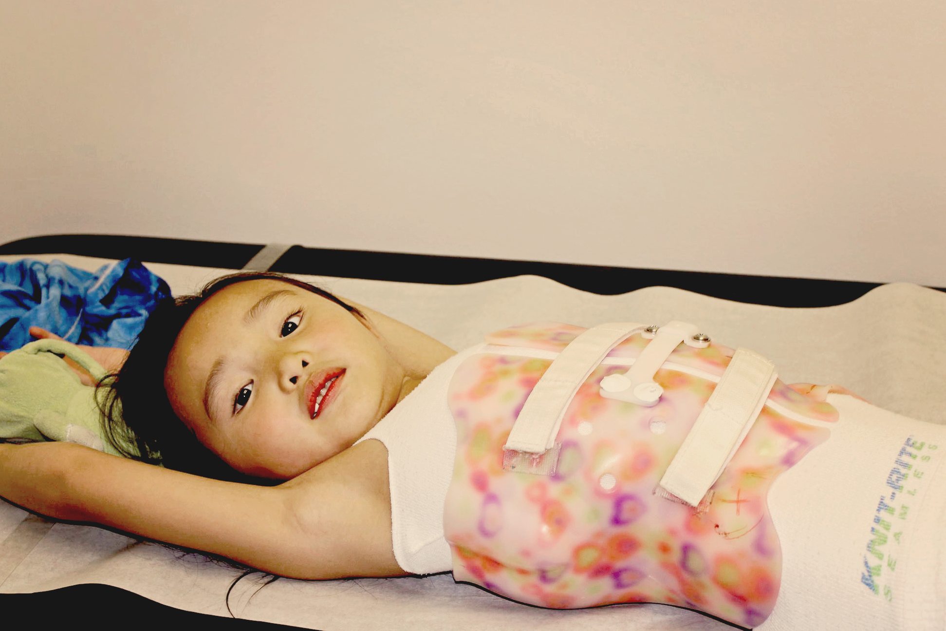
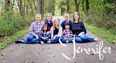
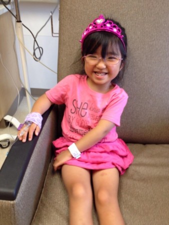
Note from Jen, a mother who has adopted a daughter with OI (pictured above):

“My daughter was 4.5 when I adopted her from China. I knew that she was relatively mild, but did not know the type. Her left femur was bowed enough that there was 1.5” difference between her 2 legs. On our 1 month home anniversary, her heel bumped at just the right angle and it broke. But it was a blessing. Our orthopedic surgeon was able to rod it and his expertise is bone lengthening. The leg is now completely straight and we gained back the lost leg length difference. Olivia also has DI and has had caps put on to protect some of her teeth. She lost her first tooth, and has 2 adult teeth coming in. She has responded very well to her infusions that she receives every 3 months (we receive zolandronic acid, the other drug option is pamidronate). Olivia has grown in height, grown in strength, runs, jumps, and loves to dance. She is one of the most positive and compassionate people I’ve ever met (not to mention most competitive). Give her a challenge and she will rise to the occasion!”

OI2

Resources:
OI Foundation
Nationwide Children’s Hospital: What is OI? and The Metabolic Bone Clinic
Shriners Hospitals
Kennedy Krieger Institute
Brittle Bone Society
All I Need- Osteogenesis Imperfecta
OI Parents Facebook Group
Parents of Adopted Children with OI Facebook Group


Read blog posts about Osteogenesis Imperfecta (OI) on No Hands But Ours.


Radial Dysplasia

Visit these blogs of families who have adopted children with Radial Dysplasia:

Amy Kratzer
Fried Rice and French Fries
The Ferrill Bunch
Two Sisters
Westhaven Kids
Greater Gifts

Visit these blogs of families with biological children with Radial Dysplasia:

Warkymom
My Twin Madness

09-04-12

Radial Club Hand, also known by the medical terms Radial Aplasia or Radial Dysplasia, is a congenital (present at birth) limb difference resulting from the partial or complete absence of the radius, the long bone on the thumb side of the forearm. There can be a wide range of involvement, but generally the absence of the radius causes the forearm to be shorter than normal and bowed inward. The palm of the hand is also smaller with the whole hand turned inward giving a club like appearance. Often the thumb is absent or small (hypoplastic thumb) and other fingers can be fused, locked, or missing.

Radial Aplasia/Dysplasia can occur on one side (unilateral) or both sides (bilateral). While Radial Aplasia/Dysplasia occurs often in isolation with no known cause, it is also associated with other congenital issues and a number of syndromes. Most commonly other issues involve the spine/vertebra, anus, heart, trachea, esophagus, and renal/kidney (VACTREL association.) Anemia and other blood disorders including Fanconi Anemia are also possible. The most common syndromes associated with Radial Aplasia/Dysplasia are Holt-Oram, and TAR syndrome.

While there is no way to create a “normal” arm, there are a number of different surgeries and procedures that can help improve function for individuals with Radial Aplasia/Dysplasia. Occupational therapy, stretching, and bracing can help with strength and range of motion. Not all individual are candidates for surgery, but centralization, which moves the hand over the remaining bone (the ulna) thus making the arm straighter, is a common procedure. A working thumb can be created from the index finger in a procedure called pollicization. Regardless of surgical or therapeutic intervention, children and adults with Radial Aplasia/Dysplasia live happy and productive lives and are able to function well on their own (Source 1 and 2).

DSCF8310

Sources:
Smiles for Kids
Children’s Hospital

Resources:
Medscape
Children’s Hospital
Smiles for Kids
Shriner’s Hospitals
Lucky Fin Project
Children with Radial Aplasia Facebook Group
Children Having Infant Limb Deficiency-CHILD Facebook Group

Medical Information on Related Genetic Syndromes:
Duane Radial Ray
Holt-Oram
TAR syndrome
VACTERL association
Fanconi Anemia


Read blog posts about Radial Dysplasia on No Hands But Ours.


Scoliosis

Visit these links to read bout families who have adopted a child with scoliosis:

Solo Gratia
The Tales of Two
Cantilina Chronicles

Scoliosis is an abnormal curving of the spine. The spine is the backbone and it runs straight down the back. Everyone’s spine naturally curves a bit. But people with scoliosis have a spine that curves too much. The spine might look like the letter C or S.


scoliosis1

This is Harper who is wearing a Thoraco-Lumbo-Sacral-Orthosis (or TLSO) brace. It is also called a Boston Brace.

scoliosis2


Most of the time, the cause of scoliosis is unknown. This is called idiopathic scoliosis. It is the most common type. It is grouped by age.

• In children age 3 and younger, it is called infantile scoliosis.

• In children age 4 through 10, it is called juvenile scoliosis.

• In children age 11 through 18, it is called adolescent scoliosis.

Scoliosis most often affects girls. Some people are more likely to have curving of the spine. Curving generally gets worse during a growth spurt.

Other types of scoliosis are:

Congenital scoliosis – This type of scoliosis is present at birth. It occurs when the baby’s ribs or spine bones do not form properly.

Neuromuscular scoliosis – This type is caused by a nervous system problem that affects the muscles. Problems can include cerebral palsy, muscular dystrophy, spina bifida, and polio.

To diagnose scoliosis, the health care provider will perform a physical exam. The child will be asked to bend forward. This makes his spine easier to see, although it may be hard to see changes in the early stages of scoliosis.

 The exam may show:

• One shoulder is higher than the other

• The pelvis is tilted

X-rays of the spine are done. X-rays are important because the actual curving of the spine may be worse than what your doctor can see during an exam.

Other tests may include:

Spinal curve measurement (scoliometer screening)

MRI of the spine

Treatment options vary and depends on many things:

• The cause of scoliosis

• Where the curve is in the spine

• How big the curve is

• If the child’s body is still growing

Treatment may include bracing, casting or surgery. Many cases of scoliosis do not require treatment at all. (info adapted from Medline Plus)


This is an x ray of Joshua, who had a large curve (138 degree) when he was adopted at age 9.

scoliosis3

The picture on the left is before his spinal fusion surgery. The picture on the right is his spine after surgery.

And here is Joshua after his surgery.

scoliosis


Resources:

Scoliosis Research Society
Rocky Mountain Scoliosis and Spine
Shriner’s Hospital for Children
Medline Plus

Facebook Groups:

Congenital Scoliosis Support! Email here to join
VEPTR Kids

Read blog posts about Scoliosis on No Hands But Ours.


Torticollis

image

Congenital Muscular Torticollis is a condition in infants and young children caused by a muscle imbalance in the neck. Children with torticollis most often have a head turn preference and a head tilt preference which can be on the same side or the opposite side. Many cases of torticollis are caused by intrauterine positioning, especially in twins. Other cases of torticollis are caused during the birth process and may result in a small lump in the neck muscle.

Early intervention is key in treating congenital muscular torticollis. Your child’s doctor should refer you for a Physical Therapy evaluation with a pediatric Physical Therapist. At the evaluation, your therapist will assess your child’s neck motion and strength, motor skills, symmetry, and visual skills. Your therapist will instruct you in exercises, positioning guidelines, and activities to promote age appropriate and symmetrical motor skills.

Infants with torticollis are at risk for a condition called Positional Plagiocephaly. This long name means a flattening of the skull bones, usually on one side more than the other. For example, a child with an untreated left head turn preference is at risk of developing a flat spot on the back of their head on the left side. There are associated facial asymmetries with Positional Plagiocephaly including prominence of the forehead on one side, one ear shifting forward on the turn side, eye asymmetry, and jaw asymmetry.

Torticollis and Plagiocephaly spiral together: a child with an untreated head turn preference will develop a flattening on one side of their head which then makes it more difficult for the child to turn their head to the “hard” side. Keeping their head turned the “easy” way then increases the flattening on that side. Promoting Tummy Time when your child is awake is a major part of treatment for torticollis and positional plagiocephaly.

Resources:
Torticollis Kids
Rainbow Kids
Identification and Treatment of Congenital Muscular Torticollis in Infants
Children’s Hospital
Gillette Children’s

Tummy Time Resources
Tummy time video
Infant Head Shape
Baby Center
Tummy Time Tips


© 2024 No Hands But Ours

The content found on the No Hands But Ours website is not approved, endorsed, curated or edited by medical professionals. Consult a doctor with expertise in the special needs of interest to you.