Vascular


Capillary Malformation (Port Wine Stain)

Visit these blogs to read about families who have adopted children with this special need:

Fireworks and Fireflies

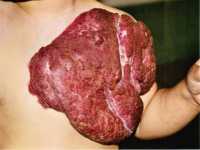
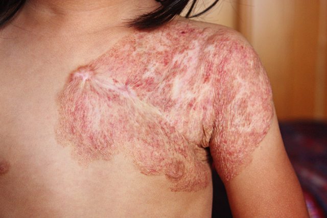
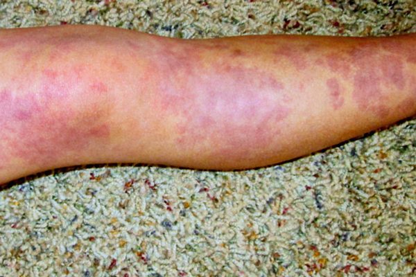
Capillary malformations, commonly known as port wine stains, occur in about 0.3% of live births. These flat, well defined areas range from pale pink to dark purple and are present at birth. Port wine stains may occur anywhere on the body, but they are predominantly found on the face and neck.


pws


The discoloration is caused by enlarged capillaries in the skin that result in increased blood flow. As capillary vessels are close to the surface of the skin, the increased blood flow gives the skin in affected areas a pink to purple appearance. Often a port wine stain is considered mostly cosmetic. However, over time the vessels can give a lumpy appearance to the skin. Also, if the stain covers an eye, or touches the nose or lip, this cobbling or thickening of the skin may impair vision and breathing in adulthood if the stain is left untreated.


Resources:
Cincinnati Children’s
Nova News
Vascular Birthmarks Foundation
Boston Children’s

Read blog posts about Port Wine Stain on No Hands But Ours.


Congenital Hemangioma

Visit these blogs to read about families who have adopted children with this special need:

Life is But a Breath

A congenital hemangioma is fully developed at birth. These hemangiomas are non-cancerous tumors composed of both thickened skin and many thin-walled blood vessels. They are most often found in the skin and are usually close to the surface though they can also be found in the liver. Typically congenital hemangiomas will present in a round or oval shape and range from pink to bluish in color, often with a grey “halo” surrounding them. These birthmarks are raised above the surrounding skin and feel warmer to the touch.


hemangioma


hemangioma2


Congenital hemangiomas are separated into two basic categories:

Noninvoluting congenital hemangiomas (NICH) and Rapidly Involuting congenital hemangiomas (RICH)

A NICH is present at birth and will grow with your child. They do not decrease in size over time. While most NICHs do not cause serious harm, you should contact a vascular anomalies specialist if a scab develops, it is on your child’s face, it is large, growing rapidly or in a spot where it could cause complications. Multiple hemangiomas may point to a hemangioma in an internal organ, and hemangiomas along the spine may point to spine issues, so these should also be checked carefully by someone experienced with vascular anomalies.

A RICH decreases in size during your child’s first year of life. While they may be completely gone by 12 to 18 months, surgery may be necessary to reduce loose skin or scarring.


hemangioma3


Resources:

Cincinnati Children’s
Children’s Hospital of Wisconsin
Nova News
Boston Children’s

Read blog posts about Hemangiomas on No Hands But Ours.


Infantile Hemangioma

Infantile hemangiomas typically present around the first two weeks of life though sometimes they aren’t visible until about four months. They may occur anywhere on the surface of the skin, but most commonly are found on the neck, face or scalp. These are the most common types of tumors (noncancerous) found in childhood, occurring in around 10% of infants. Unlike other types of birthmarks, these will grow and change quickly during the first few months. After infantile hemangiomas complete the growth phase, they begin to suddenly shrink, known as involution. This process varies and may take one to ten years to complete. Once it has involuted, it will never grow back.

Infantile hemangiomas are broken into three basic types:

Superficial (also known as Strawberry Hemangioma)
Vascular malformation.
Red, soft, raised appearance.
Size varies.
May be present at birth or first few weeks thereafter.
Will grow, but start to fade (involute), turn grey in color.
Usually disappear between ages 5-10.
Surgery might be necessary to remove – depending on size and location of lesion.
Other treatments – compression and massage, steroids, X-ray therapy, laser therapy, cryotherapy, or injection of hardening agents.

Deep (also known as Cavernous Hemangioma)
Bluish or bluish-red in color.
Lumpy mass.
Borders not visible as with other hemangiomas.
Grows fast during first 6 months – then slows – 95% disappear by 10-12 years of age.
Treatments are the same as for strawberry hemangioma.

Mixed
Most common type.
Involve both superficial and deep components.

Resources:

Boston Children’s
Nova News
Cincinnati Children’s
Children’s Hospital of Wisconsin

Read blog posts about Hemangiomas on No Hands But Ours.


Klippel-Trenaunay Syndrome

Visit these blogs to read about families who have adopted children with this special need:

coming soon!

What is Klippel-Trenaunay Syndrome?

Klippel-Trenaunay syndrome is a rare congenital (present at birth) vascular anomaly that results in your child having a large number of abnormal blood vessels. Klippel-Trenaunay syndrome is a complicated condition, and it affects each child in different ways. KTS often has three characteristic signs:

— Capillary malformation covering one or more limbs (arm or leg)

— Hypertrophy ( excessive growth of a limb) OR atrophy( withering or smaller limb)

— Abnormal blood vessels, including veins, capillaries and lymphatic vessels (vessels that drain fluid that leaks from arteries and veins and return that fluid to the bloodstream)


KTS


What causes Klippel-Trenaunay and who gets it?

No one knows the precise cause of KTS. Some doctors think it’s caused by a mutation that occurs before birth (but not one that’s inherited) in the cells that form the lymphatic system, veins and other tissues. No known food, medication or activity during pregnancy can cause Klippel-Trenaunay.

Is there a treatment for KTS?

Because there is no cure for KTS and it’s a progressive condition it is believed that treating your child’s Klippel-Trenaunay syndrome symptoms is the most effective way to manage the disease.

The first step is to have your child evaluated by members of an experienced interdisciplinary vascular anomalies team. This team may include: Dermatology, Hematology, Orthopedics, Interventional Radiology and setting up a supportive care plan.


kts


Resources:

Sturge-Weber.org
Vascular Birthmarks Foundation
RareDiseases.org

Read blog posts about Klippel-Trenaunay Syndrome on No Hands But Ours.


Parkes Weber Syndrome

What is Parkes Weber Syndrome?

Parkes Weber Syndrome is a rare, congenital (present at birth) condition that results in your child having a large number of abnormal blood vessels. It’s very similar to the more common Klippel–Trenaunay Syndrome and is treated in much the same way. But in contrast to children who have Klippel-Trenaunay syndrome, kids with Parkes Weber have arteriovenous malformations that can cause heart failure.

From the Sturge-Weber Foundation: “Parkes Weber Syndrome is characterized by one or more high-flow arteriovenous malformations leading to extremity overgrowth similar to but usually more severe than with KT. KT does not include AV malformations. An angiogram will be normal in KT and abnormal in Parkes Weber.”

What are the complications?

Possible complications of Parkes Weber include:

* Swelling and overgrowth of one of your child’s limbs (usually a leg)
* Bleeding from the capillary malformation (sometimes called port-wine birthmark)
* Heart issues due to the large amount of blood flowing to the affected limb

What causes Parkes Weber? And who gets it?

It can be hard to find accurate information for Parkes Weber as it is such a rare condition. No one knows the exact cause but some doctors believe it is caused by a mutation (in the embryonic stage of life) in the cells that form the vascular system and other tissues.

It is equally common in boys and girls.

What is the treatment?

Because there is no cure for Parkes Weber and it’s a progressive condition and this means it will grow as your child grows. It is believed that the best way to manage the syndrome is to manage the symptoms. The first step is to have your child evaluated by members of an experienced interdisciplinary vascular anomalies team. This team may include: Dermatology, Cardiologist, Orthopedics, Interventional Radiology, and setting up a supportive care plan.


Sturge-Weber Syndrome

What is Sturge-Weber Syndrome?

Sturge-Weber Syndrome( also known as SWS) is congenital, non-familial disorder of unknown incidence and cause. It is characterized by congenital facial birthmark and neurological abnormalities. Other symptoms can include: Angioma (benign tumors) on the surface of the brain leading to seizures, hemiparesis (weakness or paralysis on one side of the body) and developmental delays and glaucoma (damage to the optic nerve) on the side of the port wine stain.


corina2


What causes Sturge-Weber and who gets it?

SWS is caused by a gene acting abnormally. SWS is rare, but doctors don’t know how many people it affects. It does not run in families (inherited).

What is the treatment?

Because Sturge -Weber affects each child differenty, they should be evaluated by a team of specialist to help limit and prevent complications and to improve your child’s quality of life. This team may include:

Dermatology: Laser therapy used to lighten or diminish the port wine birthmark
Neurology: Anticonvulsant medication to control and combat seizures
Neurosurgery: To remove or disconnect the affected part of the brain when anticonvulsant medications do not prove effective
Ophthalmology: Eye drops used to treat less severe cases and surgery may be performed on individuals with more serious cases of glaucoma
Physical Therapy: For paralysis or weakness


Resources:
Sturge-Weber Foundation
Vascular Birthmarks Foundation
National Organization for Rare Disorders

Read blog posts about Sturge-Weber Syndrome on No Hands But Ours.


© 2024 No Hands But Ours

The content found on the No Hands But Ours website is not approved, endorsed, curated or edited by medical professionals. Consult a doctor with expertise in the special needs of interest to you.